A General Theory Of Magnetic Resonance Saturation: Essential
Magnetic resonance saturation occurs when a system is exposed to too much radiofrequency energy, causing it to absorb energy until it can’t anymore, disrupting the normal signal. Understanding this is essential for clear MRI scans.
Understanding Magnetic Resonance Saturation: A Beginner’s Guide
Ever feel like you’re shouting into a void, and no one’s listening? That’s a bit like what happens in Magnetic Resonance Imaging (MRI) when saturation occurs. It’s a common issue that can make your images fuzzy or unclear, and honestly, it can be a bit frustrating when you expect a crystal-clear picture. But don’t worry, it’s a normal part of how MRIs work! We’ll break down this “saturation” concept in simple terms. Think of it as understanding the limits of what your car’s engine can handle when you push it too hard. With this guide, you’ll get a clear picture of what magnetic resonance saturation is and why it matters, paving the way for better understanding of MRI technology.
What is Magnetic Resonance Saturation?
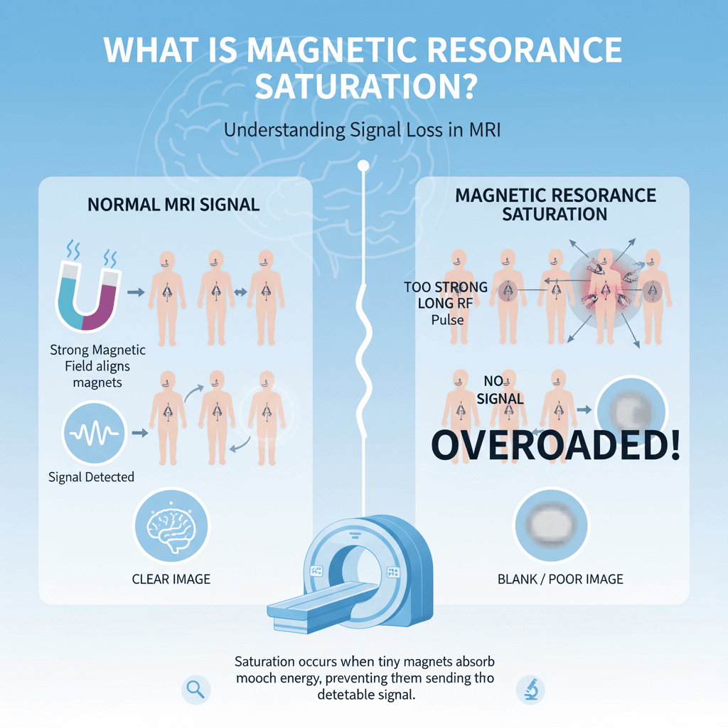
Imagine you have a bunch of tiny magnets, like little compass needles, inside your body. In an MRI machine, a strong magnetic field lines them up. Then, a radio wave pulse is sent in to give them a nudge, making them flip. As they flip back, they send out a tiny signal that the MRI machine detects to create an image.
Saturation happens when this radio wave pulse is too strong or lasts too long. It’s like giving those tiny magnets a push that’s so powerful, they get “stuck” in their flipped position, or they absorb so much energy that they can’t properly relax and send out a signal anymore. They’re essentially overloaded.
Why Does Saturation Matter?
When magnetic resonance saturation happens, the signals from those overloaded “magnets” become weak or disappear entirely. This means the MRI machine can’t pick up a clear signal from those areas. The result? Dark spots, fuzzy areas, or a lack of detail in your MRI scan. It’s like trying to listen to a quiet conversation in a stadium where everyone is cheering – you just can’t hear the important details.
For medical professionals, understanding saturation is crucial for adjusting MRI settings to get the best possible images for diagnosis. For anyone curious about how MRI works, it’s a key concept to grasp. It helps explain why sometimes scans look different and how the technology is fine-tuned.
The “General Theory” Explained Simply
The “general theory of magnetic resonance saturation” is the scientific explanation of what happens to those tiny body magnets (protons) when they’re hit with too much radiofrequency (RF) energy.
In simple terms, every proton in your body has a specific way it likes to spin and align itself in a magnetic field. When the MRI machine applies a radio wave pulse, it’s designed to “tip” these protons over. The energy of the radio wave pulse is measured in RF power.
When this RF power is just right, the protons tip and then relax back, releasing energy as a signal. But if the RF power is too high, or if the pulse is applied for too long, the protons absorb more energy than they can handle. They get “saturated.”
Tipped and Stuck: They might get tipped too far, reaching a point where they don’t naturally flip back properly.
Energy Overload: They absorb so much energy that their ability to send a reliable signal is compromised.
Relaxation Disrupted: The normal process of relaxation, which is vital for generating the MRI signal, is disturbed.
This is why controlling the strength and duration of RF pulses is so important in MRI. It’s a balancing act to get the protons to signal without overwhelming them.
Factors Contributing to Saturation
Several things can lead to magnetic resonance saturation:
High RF Input Power: The most direct cause. If the RF pulse is too strong, it can saturate the protons.
Long RF Pulse Duration: Even if the power isn’t excessively high, a pulse that lasts too long can also lead to saturation.
Repetition Rate of RF Pulses (TR): In MRI, pulses are repeated. If they are repeated too quickly (short TR), the protons don’t have enough time to fully relax between pulses. This can lead to a build-up of saturation effects.
Pulse Sequence Design: Certain advanced MRI techniques use specific patterns of RF pulses. If these are not designed carefully, they can inadvertently cause saturation.
Patient Body Habitus: Larger patients, for instance, might require higher RF power to achieve signal penetration, which can increase the risk of saturation in certain tissues.
Visualizing Saturation: An Analogy
Let’s use a simple visual analogy to understand this better.
Imagine you’re trying to fill a bucket with water.
Normal MRI: This is like gently pouring water into the bucket. The water level rises steadily, and you can see exactly how much is in there.
Saturation: This is like turning the hose on full blast directly into the bucket. Too much water comes in too fast.
Overflow: The water spills over the sides – this is like the protons not being able to send a clear signal.
Turbulence: The water inside is all churned up and not settling – a bit like the protons being in a disordered, unreadable state.
The goal in MRI is to “fill the bucket” (get a good signal) without making it overflow (saturate).
What Happens During Saturation?
When saturation occurs, the physics inside your body during an MRI scan changes in a specific way related to the longitudinal magnetization.
In a nutshell:
1. Alignment: Normally, when you’re not being pulsed with RF energy, the protons in your body align with the MRI’s main magnetic field. This alignment is called longitudinal magnetization. This is the “battery” that the RF pulse “tips.”
2. The RF Pulse: The radiofrequency pulse is designed to “tip” this alignment. Think of it like gently pushing a spinning top so it leans over.
3. Relaxation: After the pulse, the protons “relax” back to their aligned state, releasing energy. This relaxation process has two components: T1 relaxation (restoring longitudinal magnetization) and T2 relaxation (loss of signal due to dephasing).
4. Saturation Effect: If the RF pulse is too strong or too long, it tips the protons too much. Crucially, it can disrupt or completely deplete the longitudinal magnetization. This means the “battery” that powers the signal is drained. So, even if relaxation is happening, there’s not enough “stored energy” to create a strong signal.
5. Signal Loss: With depleted longitudinal magnetization after the RF pulse, the subsequent signal generated from the protons is weaker or absent. This is what saturation looks like on an MRI image – areas without a clear signal.
This disruption of the longitudinal magnetization recovery is the core of magnetic resonance saturation. It’s why understanding the pulse sequence and RF power is so vital for MRI technologists.
Key Terms to Know
Here are some important terms you might hear when discussing MRI and saturation:
Protons: Tiny particles in your body’s water and fat molecules that act like tiny magnets.
Magnetic Field: The strong, static field produced by the MRI machine that aligns the protons.
Radiofrequency (RF) Pulse: A burst of energy sent by the MRI machine to “tip” the aligned protons.
Longitudinal Magnetization: The alignment of protons parallel to the main magnetic field. This is the “signal source” that RF pulses perturb.
Transverse Magnetization: The component of magnetization that rotates in the plane perpendicular to the main magnetic field. This is what generates the detectable MRI signal.
Relaxation: The process by which protons return to their equilibrium state after an RF pulse.
T1 Relaxation: The recovery of longitudinal magnetization.
T2 Relaxation: The decay of transverse magnetization (signal loss).
Flip Angle: The angle by which the RF pulse tips the protons’ magnetization away from the longitudinal axis. A 90-degree flip angle tips it completely into the transverse plane. Higher flip angles increase the risk of saturation if not managed.
Pulse Sequence: The specific order and timing of RF pulses and gradient magnetic fields used to acquire MRI data. Different sequences have different sensitivities to saturation.
How Saturation Affects Image Quality
Saturation can manifest in several ways on an MRI scan:
Signal Loss: Areas of saturation will appear dark on the image because no signal is generated.
Uneven Brightness: You might see areas that are much dimmer than expected, indicating partial saturation.
Artifacts: In some cases, saturation can create unusual patterns or distortions in the image.
Reduced Contrast: The difference between different tissues might be blurred if saturation affects some tissues more than others.
This is why MRI technologists carefully choose pulse sequences and adjust parameters like RF power to minimize saturation in critical areas.
Saturation in Different MRI Sequences
Not all MRI sequences are equally affected by saturation. Some sequences are designed to work with or around it, while others are more susceptible.
Spin Echo (SE) Sequences: These are generally less prone to saturation than gradient echo sequences, especially with longer repetition times (TR). This is because they use a refocusing pulse to recover some signal lost due to dephasing.
Gradient Echo (GRE) Sequences: These are more sensitive to saturation. Because they use shorter TRs and smaller flip angles, the longitudinal magnetization may not have time to fully recover between pulses, leading to saturation effects.
Inversion Recovery (IR) Sequences: These sequences are specifically designed to suppress certain tissues or highlight others by using an initial 180-degree inversion pulse followed by a delay and then a standard imaging pulse sequence. If timed incorrectly, they too can be affected by saturation of the preceding pulses.
Understanding which sequence is being used helps in appreciating the potential for saturation artifacts.
Real-World Examples of Saturation
Where might you see the effects of saturation?
Near Conductive Materials: If a patient has metal implants or other conductive materials, these can interact with RF pulses non-linearly, leading to localized saturation. Some implants are safe for MRI, while others are not. For example, the U.S. Food and Drug Administration (FDA) provides guidance on MRI safety for medical devices.
High Fat or Water Content Tissues: Tissues with very high water or fat concentrations can sometimes exhibit different saturation characteristics, requiring specific sequence adjustments.
Rapid Imaging Techniques: When MRI scans are performed very quickly (e.g., for cardiac imaging or functional MRI), TR values are often shortened, increasing the likelihood of saturation.
Preventing and Managing Saturation
MRI technologists use various strategies to prevent or minimize saturation:
Adjusting RF Power: Carefully controlling the intensity of the RF pulses.
Optimizing TR: Using appropriate repetition times to allow for sufficient magnetization recovery.
Choosing the Right Pulse Sequence: Selecting a sequence that is less susceptible to saturation for the specific diagnostic need.
Using RF Shimming: A technique that aims to make the RF field more uniform across the scanned area, ensuring consistent RF delivery.
Considering Patient Factors: Adjusting scan parameters based on the patient’s size and the specific anatomy being imaged.
Tools and Technologies for Better Imaging
Modern MRI machines are sophisticated. They employ:
Advanced Pulse Design Software: Allows for precise control over RF pulse shapes and timings.
Real-time Feedback Mechanisms: Some systems can monitor signal quality and adjust parameters on the fly.
RF Coil Technology: Specialized coils (the part of the scanner that sends and receives RF signals) can help deliver RF energy more efficiently and uniformly.
These advancements are crucial for obtaining high-quality images while minimizing issues like saturation. The European Society of Radiology https://myesr.org/ often publishes research and guidelines related to advancements in imaging technology, including RF pulse design.

A Table of RF Pulse Effects
Let’s summarize how RF pulse characteristics relate to saturation:
| RF Pulse Characteristic | Effect on Longitudinal Magnetization | Likelihood of Saturation |
|---|---|---|
| High Power | Rapid and extensive tipping, depletes magnetization | High |
| Low Power | Minimal tipping, magnetization largely preserved | Low |
| Long Duration | Extended tipping, can lead to saturation over time | Moderate to High |
| Short Duration | Brief tipping, less likely to cause saturation | Low |
| Short Repetition Time (TR) | Insufficient time for recovery between pulses | Increased |
| Long Repetition Time (TR) | Ample time for recovery, magnetization can replenish | Decreased |
| Large Flip Angle | Significant tipping, closer to full saturation point | Increased |
| Small Flip Angle | Partial tipping, less likely to reach saturation point | Decreased |
Frequently Asked Questions (FAQ)
Q1: Is magnetic resonance saturation dangerous?
A1: No, magnetic resonance saturation itself is not dangerous. It’s a physical phenomenon that affects the quality of the MRI scan by reducing the signal intensity in certain areas. The RF power used in MRIs is carefully regulated to be safe for patients according to established guidelines, such as those from the International Electrotechnical Commission (IEC).
Q2: Can I do anything as a patient to prevent saturation?
A2: As a patient, there isn’t much you can do directly to prevent saturation, as it’s managed by the MRI technologist through scanner settings. However, cooperating with the technologist by staying still during the scan is crucial for achieving clear images.
Q3: Will saturation make my MRI results incorrect?
A3: Saturation itself doesn’t make results “incorrect” in terms of diagnosis. Instead, it can make certain parts of the image unclear or hard to interpret. The radiologist is trained to recognize and account for potential artifacts like saturation when reading your scan.
Q4: Why do some MRI scans look clearer than others?
A4: Image clarity depends on many factors, including the type of MRI scanner, the specific pulse sequence used for the scan, the skill of the technologist in optimizing settings, and the presence of any physical artifacts like saturation. Different parts of the body also require different imaging techniques.
Q5: How does saturation relate to the “power” of the MRI machine?
A5: The “power” of an MRI machine in this context often refers to the strength of its main magnetic field (measured in Tesla, or T) and the capabilities of its RF system. Higher magnetic fields can enable faster scanning and higher image resolution, but they also require careful management of RF power to avoid saturation.
Q6: Are there specific parts of the body where saturation is more common?
A6: Saturation can occur anywhere, but it might be more noticeable in areas where tissues have very different magnetic properties or where imaging parameters are pushed to their limits, such as in deep-seated organs or during very rapid imaging sequences.
Q7: Is saturation the same as noise in an MRI?
A7: No, saturation and noise are different. Noise is random interference that makes the image grainy. Saturation is a loss of signal due to the specific physics of RF pulses overwhelming the protons’ ability to generate a signal, often creating dark or indistinct areas.
Conclusion
Understanding magnetic resonance saturation is a key step in appreciating the sophisticated science behind MRI. It’s not a flaw in the system, but rather a predictable outcome when the delicate balance of radiofrequency energy is tipped too far. By learning about how RF pulses interact with your body’s magnetic properties, you gain a clearer picture of why image quality can vary and how expertly trained professionals ensure you get the best possible diagnostic information. Remember, the goal is always a clear signal, and mastering concepts like saturation is part of that intricate process. Keep learning about the amazing technology that helps us see inside ourselves!